Main Highlights

Rangers’ Corey Seager Hit by Pitch: Left Shin Contusion, Status for Next Game Unclear
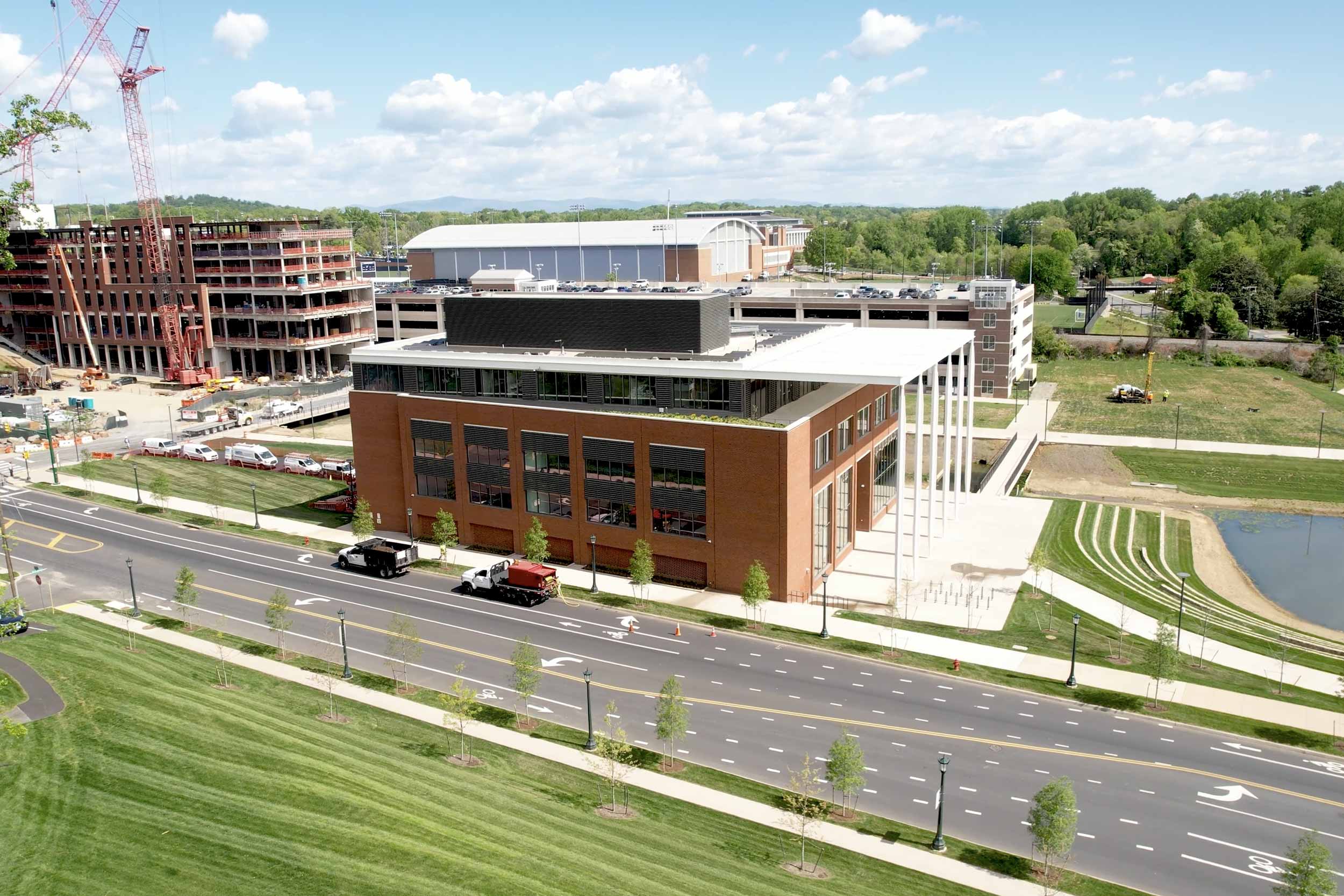
Sustainability Meets Innovation: The Construction of UVA’s School of Data Science Building
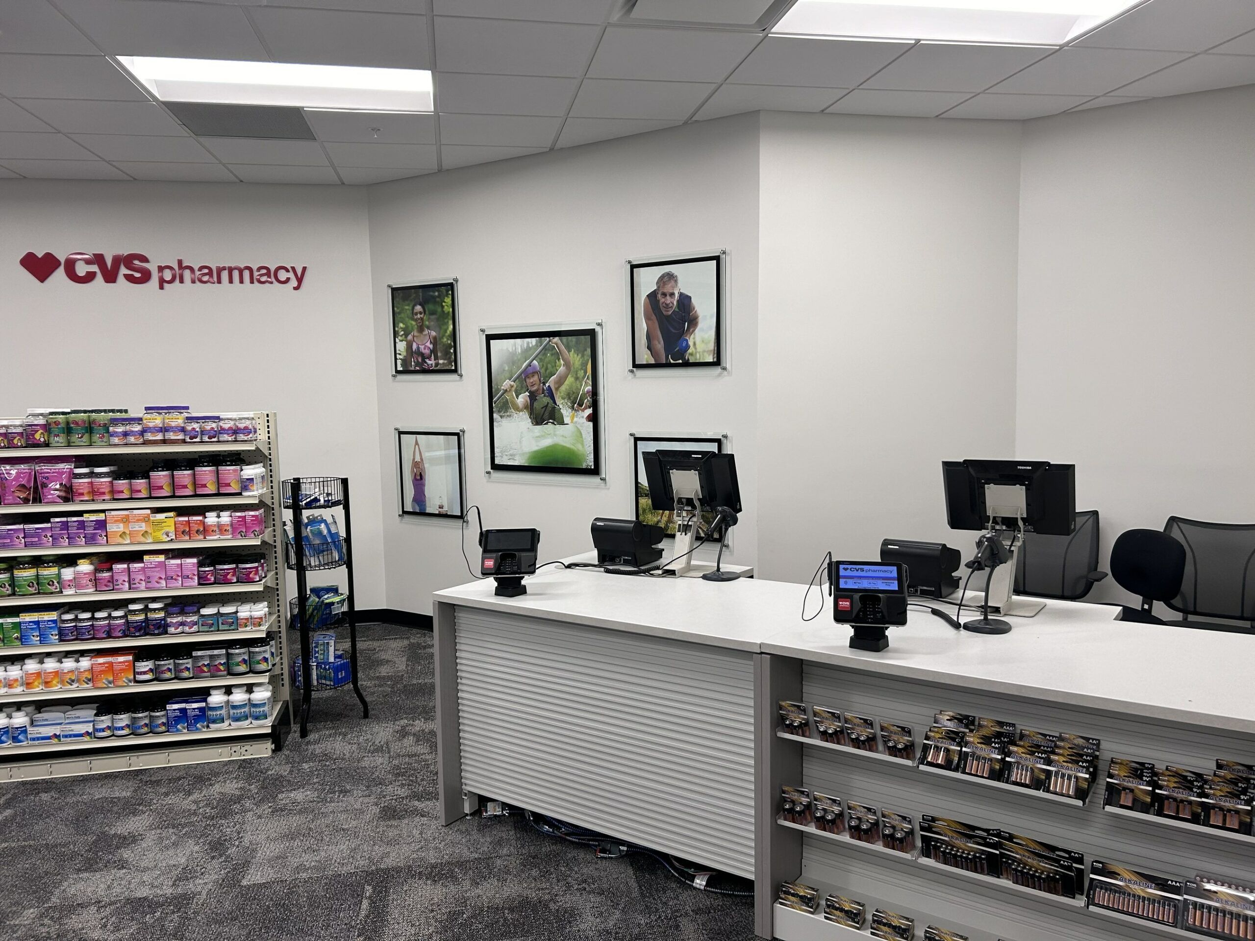
CVS Health Opens Facility in Baton Rouge to Help Community Overcome Employment Barriers and Improve Health Outcomes